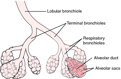

Within the lungs, the main (primary) bronchi branch into lobar (secondary) bronchi. Left main bronchus – passes inferiorly to the arch of the aorta, and anteriorly to the thoracic aorta and oesophagus in order to reach the hilum of the left lung.Clinically, this results in a higher incidence of foreign body inhalation. The right superior lobar bronchus arises before the right main bronchus enters the hilum. Right main bronchus – wider, shorter, and descends more vertically than its left-sided counterpart.Each secondary bronchi supplies a lobe of the lung, and gives rise to several segmental bronchi.Īlong with branches of the pulmonary artery and veins, the main bronchi make up the roots of the lungs. They undergo further branching to produce the secondary bronchi. The trachea receives sensory innervation from the recurrent laryngeal nerve.Īrterial supply comes from the tracheal branches of the inferior thyroid artery, while venous drainage is via the brachiocephalic, azygos and accessory hemiazygos veins.Īt the level of the sternal angle, the trachea bifurcates into the right and left main bronchi. This is the most sensitive area of the trachea for triggering the cough reflex, and can be seen on bronchoscopy. This acts to trap inhaled particles and pathogens, moving them up out of the airways to be swallowed and destroyed.Īt the bifurcation of the primary bronchi, a ridge of cartilage called the carina runs anteroposteriorly between the openings of the two bronchi. The combination of sweeping movements by the cilia and mucus from the goblet cells forms the functional mucociliary escalator. The trachea and bronchi are lined by ciliated pseudostratified columnar epithelium, interspersed by goblet cells, which produce mucus. The free ends of these rings are supported by the trachealis muscle.

The trachea, like all of the larger respiratory airways, is held open by cartilage – here in C-shaped rings. Key: Green – upper lobe, yellow – middle lobe, blue – lower lobe By Bibi Saint-Pol, via Wikimedia Commonsįig 1 – Overview of the tracheobronchial tree.


 0 kommentar(er)
